|
|
“The science of embryology and our knowledge of the evolution of man is the foundation of medicine. They are the two sources that reveal to us the nature of cancer and of all so-called diseases” (Dr. med. Ryke Geerd Hamer).
|
|
DEVELOPMENT FROM THE ORIGINAL RING FORM TO THE FINAL EMBRYO FORM
Human life begins as a single cell holding all instructions for its growth and development. Starting with the first cell division, the embryo grows into a cluster of cells called a blastocyst. Two weeks after conception, the blastocyst divides into three embryonic germ layers: an inner endoderm, an outer ectoderm, and a mesoderm forming in between. Over the course of gestation, the embryonic germ layers develop all organs and tissues of the body. Throughout this period, the growing fetus passes through all the evolutionary stages from a single-celled organism to a complete human being. The three germ layers give rise to the same tissue types in all organisms, including animals and plants.
NOTE: The theory that the development of the fetus (ontogeny) recapitulates the evolutionary history of all remote ancestors (phylogeny) was formulated in the 1800s by German biologist Ernst Haeckel. Since the early twentieth century, Haeckel's “biogenetic law” has been refuted on many fronts. Dr. Hamer's scientific work offers a new and extended reading of Haeckel's theory by showing that the evolutionary development of the human organism, including the brain, represents biological conflict themes that had once been phases in evolution. This proves that Haeckel's claim is fundamentally true.
|
|
|
We know from the science of biology that the first life forms were ring-formed organisms consisting solely of intestine. At this early development stage, the GULLET (primordial oro-pharyngo-anal cavity) served both the intake of food and the disposal of feces. The ingoing section of the intestinal canal regulated ingestion and digestion, the outgoing section elimination (see diagram).
The image on the right shows a five days old human embryo. The ring form is still maintained.
|
|
| The nerve distribution of the autonomic nervous system before birth also points to the primordial ring form. While the sympathetic nerves are arranged in the middle of the spinal cord, the parasympathetic (vagotonic) nerves are located on the periphery, namely at the base of the brain and in the sacral region, close to the pharynx and the rectum. This strongly suggests that the parasympathetic divisions were once connected.
We have to envision the development of the spinal cord and the spine progressively from the cervical (C), thoracic (T) and lumbar spine (L) to the sacrum; first, in a round configuration equal to the ring form of the intestine. We can speak of an upper and lower section of the spine only after the gullet had broken open. The sympathetic trunks, which are two long chains of nerves on each side of the vertebrae, allow nerve fibers to travel to spinal nerves that are superior or inferior to the one in which they originate.
|
|
| In the BRAINSTEM, the oldest part of the brain, the control centers of the organs of the intestinal canal are also arranged in a ring-form order, starting on the right hemisphere with the brain relays of the mouth and pharynx (incl. thyroid gland, parathyroid glands), esophagus, stomach, liver parenchyma, pancreas gland, duodenum, small intestine, continuing counter-clockwise with the brain relays of the appendix, cecum, colon, rectum and bladder on the left side of the brainstem. The transition from the right to the left brainstem hemisphere corresponds on the organ level to the ileocecal valve, positioned between the small intestine and the cecum, the first section of the large intestine.
The lung alveoli, middle ear and Eustachian tubes, tear glands, choroid, iris and ciliary body of the eyes, kidney collecting tubules, adrenal medulla, prostate, uterus and fallopian tubes, Bartholin’s glands, smegma producing glands as well as the pituitary gland, pineal gland, and choroid plexus originate from the intestinal mucosa. They are therefore controlled from the brainstem. |
|
| All organs that are controlled from the BRAINSTEM derive from the ENDODERM, the first and oldest embryonic germ layer. Because of their origin from the intestinal mucosa, they consist of INTESTINAL CYLINDER EPITHELIUM.
|
|
|
Over the course of evolution, the GULLET BROKE OPEN. The new opening of the outgoing section developed into today’s rectum, the remaining gullet became in its entirety the mouth and pharynx (see diagram).
The image on the right shows the further development of the fetus to the final embryo form, outlining the embryonic germ layers.
|
|
| The intestinal rupture occurred close to the left half of the gullet. This explains why the control center of the mouth and pharynx is divided into two brain relays located opposite each other at the midline of the brainstem hemispheres.
The right half of the mouth and pharynx is controlled from the right side of the brainstem that still regulates ingestion (“ingoing morsel”), while the left half of the mouth and pharynx is controlled from the left side of the brainstem, which, however, no longer regulates excretion (this is now managed by the rectum) but instead the vomiting reflex (a remainder of the gullet’s previous fecal disposal function). The preservation of the original innervation of the left half of the gullet also serves the biological purpose to be able to disgorge a morsel ( excretory quality) that might cause harm to the organism. |
| The rupture of the gullet happened at a point in time when so-called SQUAMOUS EPITHELIUM that originated from a new embryonic germ layer, namely from the ECTODERM, had already migrated from the gullet both into the ingoing and outgoing section of the intestine. During gestation, the ectoderm develops on the seventeenth day after fertilization. All organs and tissues that derive from the ectoderm are controlled from the CEREBRAL CORTEX. NOTE: The alpha islet cells and beta islet cells of the pancreas , the olfactory nerves, and the thalamus are controlled from the diencephalon (part of the cerebrum).
|
|
|
The starting point of the ectodermal cell migration was the squamous epithelium covering the periosteum of the paranasal sinuses. The sensitive nerves of the epithelial sinus mucosa provided a heightened sense of smell facilitating survival (scent of danger) as well as procreation (scent of a mate).
|
The squamous epithelial cell migration into the INGOING SECTION OF THE GULLET explains why ectodermal tissue is found in today's …
|
|
… esophagus (upper two-thirds), stomach (small curvature), pylorus, duodenal bulb, bile ducts, gallbladder, pancreatic ducts, coronary arteries, coronary veins, ascending aorta, internal carotid arteries, inner sections of the subclavian arteries, carotid sinus, glans penis, and glans clitoris. All these tissues are controlled from the POST-SENSORY CORTEX. |
|
|
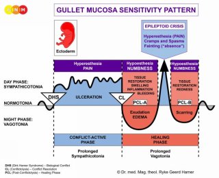
Both organ groups follow the GULLET MUCOSA SENSITIVITY PATTERN (so named because of its connection to the gullet) with hypersensitivity during the conflict-active phase and the Epileptoid Crisis and hyposensitivity during the healing phase. |
The squamous epithelial cell migration into the OUTGOING SECTION OF THE GULLET explains why ectodermal tissue is found in today’s …
|
|
NOTE: After the gullet had broken open, the sensitive nerves as well as the motor innervation of the entire urino-rectal system had to be rewired through the spinal cord (this is why these organs paralyze with paraplegia) and were connected to the OUTER SKIN SENSITIVITY PATTERN. In the brain, the organs are orderly arranged side by side on the left side of the cerebral cortex.
|
The MESODERM, which developed after life had moved on land, is divided into an older and younger group.
| The NEW MESODERM develops the bones (incl. bone marrow and blood cells), tooth dentin, periodontium, myocardium, striated muscles, cartilage, tendons, ligaments, fat tissue, connective tissue (incl. neuroglia and myelin), endocardium and heart valves, blood vessels (incl. descending aorta, external carotid artery, outer sections of the subclavian arteries, abdominal aorta, cerebral arteries), meninges, lymph vessels with lymph nodes, spleen, ovaries, testicles, corpora cavernosa (penis), kidney parenchyma, adrenal cortex, and parts of the vitreous body. All organs and tissues that derive from the new mesoderm are controlled from the CEREBRAL MEDULLA, which had formed underneath the cerebral cortex.
|
|
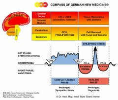
| The ability of the primordial cell to divide through mitosis, creating diploid cells which contain two sets of chromosomes, became the blueprint for Old Brain (brainstem and cerebellum) controlled organs that generate cell proliferation during the conflict-active phase. The so-called reduction division (meiosis) where the number of chromosomes is reduced from diploid to haploid became the plan for cerebrum (cerebral medulla and cerebral cortex) controlled organs that generate cell loss during conflict activity. The Biological Special Programs are inscribed in the genetic make-up of each cell of the human organism. | NOTE: Originally, these biological survival programs were directed from the “organ brain”. With the growing complexity of life forms, however, a “head brain” developed from where each Biological Special Program is coordinated. The transfer from the “organ brain” to the “head brain” explains why, in line with evolutionary reasoning, the control centers in the brain are arranged in the same order as the organs in the body.
|
|
|
NOTE: The bones of the skeletal system are supplied by the spinal nerves. The innervation of the bones comes from the second to fourth cervical nerves (C2-C4). The corium skin is supplied by the second to fifth cervical nerves (C2-C5), almost parallel to the bone innervation. The epidermis is supplied by the fifth to seventh cervical nerves (C5-C7). The reason for the different innervation of the bones and the epidermis is that the bones, originating from the new mesoderm, developed much earlier than the outer ectodermal layer of the skin.
|
At first, the periosteum that envelops the bones of the skeletal system was covered with squamous epithelium. After the muscles, ligaments, tendons and two skin layers ( corium skin and outer skin) had given new support to the bones, the squamous epithelial layer degenerated (in the fetal development this process occurs during the first two weeks of gestation). What remained was a sensitive network of periosteal nerves (controlled from the post-sensory cortex).
THE DEVELOPMENT OF MUSCLE TISSUE
SMOOTH MUSCLES: The smooth muscles of the human body originate from the intestinal muscles of the primordial oro-pharyngo-intestinal-rectal canal.
| The smooth muscles of the intestines, sigmoid colon and rectum (upper part), internal anal sphincter, renal pelvis, ureters, bladder, urethra, internal bladder sphincter, esophagus, bronchia, trachea, larynx, uterus, myocardium (atria), blood vessels (incl. coronary arteries, coronary veins, aorta, carotid arteries, subclavian arteries), lymph vessels, pupils, and the smooth ciliary muscles originate from the ENDODERM.
Smooth muscles are involuntary non-striated muscles. Their ability to contract allows moving the “ food morsel” (intestinal muscles), the “ blood morsel” (atria, blood vessels), the “ air morsel” (laryngeal muscles, bronchial muscles), the “ urine morsel” (renal pelvis, ureters, bladder, urethra, internal bladder sphincter), the “semen morsel” ( prostatic ducts), and the “ light morsel” (pupil muscles) through specific organs by peristaltic motion.
The smooth muscles are controlled from the MIDBRAIN, located at the outermost part of the brainstem. NOTE: The male and female germ cells are also controlled from the midbrain.
In the event of a biological conflict, the related muscles generate during the conflict-active phase cell proliferation with an increase of muscle mass and increased local muscle tension (hypertonus). In the healing phase, the muscles relax. The Epileptoid Crisis presents as muscle spasms. In the uterus, the additional muscle cells remain after healing has been completed. |
STRIATED MUSCLES: The striated muscles developed at a time when more efficient muscle functions were required.
| The striated muscles of the skeletal musculature, myocardium (ventricles), coronary arteries, coronary veins, aorta, carotid arteries, and subclavian arteries, blood vessels, tongue, jaw, ear, bronchial muscles, laryngeal muscles, diaphragm, esophagus, stomach (small curvature), pylorus, duodenal bulb, pancreatic ducts, bile ducts, gallbladder, cervical muscles, cervical sphincter, vaginal muscles, rectal muscles, external anal sphincter, renal pelvis, ureters, urethra, bladder muscle, external bladder sphincter, eyelid muscles, ciliary muscles, and extraocular muscles derive from the NEW MESODERM.
The trophic function of the striated muscles is controlled from the CEREBRAL MEDULLA.
The ability to move the muscles is controlled from the MOTOR CORTEX.
|
Lastly, the ECTODERM developed the OUTER SKIN that covered the entire corium skin (under skin). From the outer skin, ectodermal squamous epithelium migrated through the nipples into the milk ducts, into the ear canal, nasal cavities and respiratory tract. It also covered the outer part of the eyes. This is why squamous epithelium is found in today’s …
|
|
… epidermis (including the external genitals and the vagina), prostatic ducts, eyelid skin, eyelid gland ducts, conjunctiva, cornea, lens, milk ducts, outer ear and auditory canal, nasal mucosa, trachea, larynx and vocal cords, and bronchia. All these tissues are controlled from the SENSORY CORTEX.
|
|
|
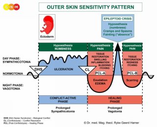
This organ group follows together with the organs of the urino-rectal system the OUTER SKIN SENSITIVITY PATTERN (so named because of its connection to the outer skin) with hyposensitivity during the conflict-active phase and the Epileptoid Crisis and hypersensitivity in the healing phase. |
|
|
This GNM diagram, showing a basal view of the cerebral cortex, illustrates that the pre-motor sensory cortex and post-sensory cortex (which control all organs following the Gullet Mucosa Sensitivity Pattern, the urino-rectal system, and the periosteal nerves) are considerably larger than the sensory cortex and motor cortex.
|
|
|
The pre-motor sensory cortex and post-sensory cortex were originally one big area that was later separated by the sensory and motor cortex maintaining a connection only at the cranial base.
|
| The retina and the vitreous body of the eyes derive from the ECTODERM. They are controlled from the VISUAL CORTEX located in the occipital lobe in the back of the brain. The visual cortex and its corresponding organs developed before the sensory and motor cortex.
|
|


