
KIDNEYS and BLADDER
 DEVELOPMENT AND FUNCTION OF THE KIDNEY COLLECTING TUBULES: The kidneys are positioned on each side of the lower spine at the back of the abdomen (retroperitoneal). The function of the kidney collecting tubules is to collect urine produced in the kidney parenchyma and funnel it through several cup-shaped calyces to the renal pelvis. From there, the urine passes further into the ureters, bladder and urethra for excretion. Urine consists for the most part of water (about 95%). The rest is made up of electrolytes (mainly sodium, potassium, chloride, and calcium) and uric substances such as uric acid, urea and creatinine. The kidneys filter daily approximately 180 liters of blood. However, 99% of the filtrate is reabsorbed by the kidney tubules and returned to the bloodstream, leaving a urine output between 1.5 and 2 liters.
In evolutionary terms, the kidney collecting tubules are the oldest tissue of the kidneys. Like the intestinal cells that digest the “food morsel”, the biological function of the kidney tubules is to “absorb/retain” (absorptive quality) and “digest” (secretory quality) the “water morsel”. The kidney collecting tubules consist of intestinal cylinder epithelium, originate from the endoderm and are controlled from the brainstem.
BIOLOGICAL CONFLICT: The biological conflict linked to the kidney collecting tubules originates at a time when life existed only in the ocean and being thrust out of the water environment created a life-threatening situation. This kind of distress also concerns human life because water is the primordial home of all living organisms. We humans experience the conflict of “feeling like a fish out of water” when we are unexpectedly “swept out” of our familiar surroundings or when we lose our “pack”. In GNM, we refer to the conflict of the kidney collecting tubules as to an abandonment conflict, existence conflict, or refugee conflict.
Abandonment conflicts are brought on by feeling ousted, excluded, unwanted, rejected, not understood, ignored, left out, isolated and alone. Children experience the conflict when they are put into daycare, when they feel unloved or excluded from the group (at home, on the playground, in kindergarten, at school), when their parents don't spend enough time with them, when a new sibling is born who gets more attention, when a grandparent dies, or when a family member leaves. It is the loss of safety and the loss of an emotional shelter which makes them feel utterly alone. The same can be said about the elderly who end up in nursing facilities, away from their home and their family. Newborns are equally vulnerable. Thus, being taken away from the mother at birth for one or the other reason can cause a severe abandonment conflict. Pets also suffer terribly when they are left behind.
An existence conflict is a fear for one's life - equal to the fish out of water in danger of dying. This fear is often triggered by a cancer diagnosis or negative prognosis associated with “my life is at stake” (compare with death-fright conflict related to the lungs). Waiting in an emergency room, being in an ambulance, and hospitalization (undergoing chemo treatments, surgery, not feeling cared for, a lack of support by doctors, nurses, or relatives) also evoke existence and abandonment conflicts. The fear of having to go to the hospital might already activate the conflict. An existence conflict also relates to one's livelihood. The feeling behind the conflict is “I have lost everything”. This could be the loss of a workplace, financial losses, the loss of a home, or the loss of a person who provided security, economically or emotionally.
A refugee conflict is experienced as being “thrown into the desert”, as feeling uprooted or “in exile”, for example, due to an unexpected transfer or move (change of neighborhood, change of school) or being forced to flee from one's home or homeland. Traveling away from a familiar home or a loved one can provoke the conflict. Air travelers are particularly prone to suffer refugee conflicts. By the same token, feeling uncomfortable on an airplane (a fear of flying) might trigger an existence conflict.
CONFLICT-ACTIVE PHASE: Starting with the DHS, during the conflict-active phase cells in the kidney tubules proliferate proportionally to the intensity of the conflict. The biological purpose of the cell increase is to close the excretion filter in order to withhold water so that the organism has a better chance to survive. This innate water retention program is vital because without water all metabolic processes stop functioning. NOTE: Whether the conflicts affect the right or left kidney is random.
Symptoms of the conflict-active phase:
The degree of WATER RETENTION is determined by the intensity of the conflict. Typical signs of water retention are baggy eyes, swollen hands, swollen feet and ankles (see also peripheral edema) and weight gain (1 liter of retained water weighs about 1 kilo or 2.2 pounds). With a persisting abandonment or existence conflict a person can gain a lot of weight (100 kg and more) in spite of regular exercises, a normal diet or even fasting. The retained water is predominantly stored in the fat tissue, mainly in the abdominal area (see ascites). In this case, obesity is not caused by excess body fat but by excess accumulation of water as a result of lasting conflict activity (compare with obesity due to hypoglycemia).
German New Medicine offers an entirely new understanding of the increasing number of overweight people, including children, in the Western world by taking into account social changes (the dissolution of traditional family structures, growing divorce rates, infants in daycare, the elderly in homes) and alarming economic developments (increasing unemployment, poor prospects for the youth, mounting debt). Whether we consider nowadays water retention (weight gain) useful or not is irrelevant. What matters is that this Biological Special Program has proved itself biologically meaningful over millions of years.
In the conflict-active phase, the organism not only withholds water but also uric substances such as uric acid, urea, and creatinine. Hence, these levels rise proportionally to the degree of conflict activity and the number of kidney tubules that are affected (compare with elevated uric acid, urea and creatinine levels related to the kidney parenchyma). The standard theory that ELEVATED URIC ACID LEVELS are linked to a diet high in proteins (see gout) is inconclusive since vegetarians also happen to have high levels of uric acid.
Urea and creatinine are waste products of the protein metabolism and are normally excreted with the urine. However, in the critical event of an existence conflict, the organism recycles the retained substances into protein to provide the organism with nutrition. Why? Because, in biological terms, the conflict of being thrust out of the water environment means next to the danger of drying out also a threat of starvation, particularly of dying from protein deficiency. For this emergency situation Nature created yet another survival program, which is to convert toxins such as urea and creatinine into food to help the organism to overcome the crisis. ELEVATED UREA AND CREATININE LEVELS are therefore not diseases (“uremia”) or malfunctions (“kidney insufficiency”), as claimed by conventional medicine, but serve a biological purpose. The retention of urea and creatinine is in addition to storing water an innate response in case water and protein are not available for a longer period of time.
The retention of water and urine results in a DECREASED URINE OUTPUT. Thus, during the conflict-active phase the urine is concentrated and dark yellow. Since water is also absorbed from the intestines, the stool is dry and hard. When more kidney tubules are involved, the urine excretion can decrease drastically causing oliguria (urine output between 150-400 ml daily) or anuria (less than 50 ml per day).
With prolonged conflict activity a flat (absorptive type) or cauliflower-shaped growth (secretory type) develops in the kidney collecting tubules. In conventional medicine, this is diagnosed as a kidney cancer or “renal cell carcinoma” (compare with “kidney cancer” related to the kidney parenchyma). If the rate of cell division exceeds a certain limit, the cancer is considered as “malignant”.
CONFLICT RESOLUTION: With the resolution of the conflict (CL) the retained water is immediately released through the unaffected calyces. Depending on the degree of water retention the elimination of urine could be profuse. Standard medicine views this copious urination (polyuria) as “abnormal” and “pathological”. With the knowledge of GNM, we welcome this URINARY PHASE with great relief (see also urinary phase shortly after every Epileptoid Crisis).
HEALING PHASE: Following the conflict resolution, fungi or mycobacteria such as TB bacteria remove the cells that are no longer required. Healing symptoms are a cloudy urine since the discharge produced during the decomposing process is excreted via the urinary tract (the discharge might contain blood), pain due to the swelling, and night sweats. With an inflammation the condition is called “nephritis” (compare with glomerulonephritis related to the kidney parenchyma). Renal candidiasis reveals that fungi assist healing.
If TB bacteria are present this causes a “bacterial kidney infection” or kidney tuberculosis (compare with “kidney infection” related to the renal pelvis, see also “bacterial kidney infection” involving E.coli bacteria). After the tuberculosis, particularly when the healing phase lasted for a long period of time, the affected calyces appear on an X-ray plump rather than with sharp contours. It is from this roentgenological appearance that doctors make the diagnosis “nephrotic syndrome” (see also renaming of lung tuberculosis to lung cancer and liver tuberculosis to liver cancer).
Tubercular secretion is rich in protein. Hence, when the additional cells are broken down, the elimination of protein through the urine is higher than normal. This is clinically termed proteinuria or albuminuria (in conventional medicine, protein in the urine during pregnancy is considered a “pregnancy disorder”, called pre-eclampsia). In the blood, however, the protein concentration is low (hypoproteinemia) because, in the event of a protein deficiency, the organism takes proteins from the blood in order to balance the protein loss. If protein-rich nutrition or supplementation is not sufficient to correct a protein shortage, administering albumin infusions temporarily is crucial. At the end of the healing phase, the protein levels as well as the urea and creatinine values are back to normal.
In the event of a new kidney tubules-related conflict, a cirrhotic kidney is no longer able to retain water. As a consequence, large volumes of diluted urine are eliminated. This condition is called diabetes insipidus. The theory that diabetes insipidus is linked to a “hormonal defect” is pure assumption.
When the affected kidney is surgically removed, a new or re-activated abandonment or existence conflict affects the other kidney because the water retention program has highest priority. This initiates the development of a new kidney tumor, interpreted by conventional medicine to be a “metastatic cancer”.
If the required microbes are not available upon the resolution of the conflict, because they were destroyed through an overuse of antibiotics, the additional cells remain. Eventually, the growth becomes encapsulated. In the kidney, this could cause an occlusion of the opening to the renal pelvis. In this case, surgery might have to be considered.
KIDNEY STONES (Calcium Oxalate Stones)
With constant conflict relapses the accumulating salt and mineral residues in the kidney collecting tubules eventually form kidney stones, which are released during the Epileptoid Crisis with spasms (kidney colic) and acute pain, particularly if a stone obstructs the urinary tract (see also kidney colic related to the renal pelvis).
|


THE KIDNEY COLLECTING TUBULES SYNDROME
 The Kidney Collecting Tubules Syndrome, in short: the SYNDROME, involves:
When the organism withholds water, the excess fluid is also stored in the healing organ and in the correlating brain relay. Hence, the size of the edema that develops in PCL-A (exudative phase) is not only determined by the magnitude of the conflict and the intensity of the preceding conflict-active phase but also by the degree of water retention due to an active abandonment or existence conflict. Whether water retention is responsible for large swellings in the healing phase can easily be established by evaluating the urea and creatinine levels and by measuring the urine output. In the practical application of GNM, a brain CT analysis is an invaluable diagnostic tool for assessing the situation.
The SYNDROME can create serious complications both on the organ and the brain level, specifically during the Epileptoid Crisis.
Consequences of the SYNDROME on the ORGAN LEVEL
The most dramatic healing phases occur therefore with the SYNDROME, that is, with concurrent water retention.
Consequences of the SYNDROME on the BRAIN LEVEL
MEDICATION with the SYNDROME
In general, all medications with a stimulating effect, including cortisone, cytostatic drugs, and morphine, exacerbate the symptoms of the conflict-active phase. In case of an abandonment or existence conflict involving the kidney collecting tubules they, therefore, increase the water retention. As a result, swellings (edemas) that occur in the first part of the healing phase (PCL-A) become much larger!
Cortisone stimulates the sympathetic nervous system. This is why it reduces vagotonic symptoms such as inflammations and swellings (the same applies to topical steroid creams). After discontinuation of the treatment, the healing symptoms therefore quickly return. Hence, administered during the healing phase, the drug only interrupts the healing process. From a GNM point of view, cortisone is only recommended in the exceptional case of a large brain edema for the purpose to reduce the brain pressure before the onset of the Epileptoid Crisis. However, according to Dr. Hamer, with the SYNDROME corticosteroids are contraindicated since they increase the water retention resulting in enlarged swellings, which can lead to life-threatening complications.
Cytostatics are highly poisonous drugs that inhibit cellular growth. In conventional medicine they are employed to “kill cancer cells”. Based on the knowledge of the Five Biological Laws and the understanding that the cell proliferation (“cancer”) has a biological purpose in the conflict-active phase and a restorative function in the healing phase, chemo drugs, including methotrexate, severely disrupt the natural course of a Biological Special Program (“disease”). Next to their toxicity, cytostatics have a highly stimulating effect. Hence, with an active existence conflict, often triggered by the cancer diagnosis itself, tumors enlarge drastically due to the increased water retention. Ironically, this is then interpreted as a “fast growing” and “aggressive” cancer. The low urine output (at this point called “kidney insufficiency”) also prevents toxins from being sufficiently eliminated. Moreover, chemo treatments weaken the elasticity of the brain tissue that undergoes healing. Eventually, the brain tissue ruptures causing death. Cytostatics suppress the production of blood cells which is devastating in the treatment of leukemia.
Morphine is known as a narcotic pain reliever. It also activates the hormone ADH (antidiuretic hormone) limiting the formation of urine. Together with its stimulating properties the drug increases the water retention and therefore the swelling both on the organ level and the brain level. With the SYNDROME morphine affects the brain much the same way as chemo treatments (cytostatics). In addition, morphine paralyzes the intestines with the result that food can no longer be processed; it also thins the lung tissue making it prone to rupture. When the effect of the drug wears off, the person falls into a state of deep vagotonia and potentially into a coma. Morphine is an opium derivative (see also codeine), hence, the sedating effect. In today’s medicine it is given to patients to “pass over easier”.
 In the majority of cases the SYNDROME is caused by a diagnosis shock, the fear associated with the “disease”, and hospitalization. Hence, resolving the kidney tubules-related conflict must have absolute priority. The resolution of the existence conflict initiates the instant release of the retained water (urinary phase) with the effect that the swellings go down quickly. This can be life-saving!
If the conflict cannot be resolved at the time, Dr. Hamer recommends salt baths with a salt concentration of 0.9% (1 kg of salt to 99 liters of water) in order to address the “fish out of water”-conflict on a solely biological level. By taking the organism “home” to the sea, the body is able to eliminate large amounts of urine. In addition, isotonic salt baths balance the salt content of blood serum, which decreases during the healing phase of the kidney tubules due to the protein loss.
Diuretics (water pills) should only be considered an emergency measure since their sympathicotonic properties actually increase the water retention while forcing urination at the same time; hence, their “side effects” on the kidneys. In addition, diuretics only eliminate electrolytes but not uric substances such as urea and creatinine. However, if diuretics are taken together with sodium bicarbonate, better known as baking soda, the kidneys will excrete uric substances in sufficient amounts. The reason for this is that sodium bicarbonate increases the glomerular filtration rate (the rate at which the kidneys filter blood). This means that an increased amount of glomerular filtrate gets into the kidney collecting tubules, which in turn increases the volume of urine.
As documented by Homer W. Smith in From Fish to Philosopher, sodium bicarbonate is a salt that was abundantly present in the primordial ocean. When life was leaving the water environment, sodium bicarbonate was absorbed into the bloodstream to be prepared for living and surviving on land. In the human organism sodium bicarbonate also plays a significant role in maintaining the acid-base balance.
|


 DEVELOPMENT AND FUNCTION OF THE ADRENAL MEDULLA: The adrenals are paired hormonal glands seated on top of the kidneys. The adrenal medulla, at the core of the gland and surrounded by the adrenal cortex, consists of so-called chromaffin cells, named for their characteristic brown staining with chromic acid salts. The adrenal medulla produces hormones (hormonal quality); predominantly stress hormones such as dopamine, noradrenaline, and adrenaline (also known as catecholamines). The adrenal medulla consists of intestinal cylinder epithelium, originates from the endoderm and is therefore controlled from the brainstem.
BIOLOGICAL CONFLICT: unbearable intense stress
CONFLICT-ACTIVE PHASE: Starting with the DHS, during the conflict-active phase adrenal cells proliferate proportionally to the intensity of the conflict. The biological purpose of the cell increase is to enhance the production of stress hormones in order to improve the performance during acute stress. Hence, the dopamine, noradrenaline and adrenaline levels rise. Symptoms are onsets of rapid heartbeats, elevated blood pressure, excessive sweating, and anxiety due to the intense state of stress. NOTE: These parameters increase to a certain extent in the conflict-active phase of any Biological Special Program.
With lasting conflict activity a compact, cauliflower-shaped growth (secretory quality), referred to as an adrenal cancer (pheochromocytoma), develops in the adrenal medulla (compare with “adrenal cancer” related to the adrenal cortex). If the rate of cell division exceeds a certain limit, conventional medicine considers the cancer as “malignant”.
HEALING PHASE: Following the conflict resolution (CL), fungi or mycobacteria such as TB bacteria remove the cells that are no longer needed. Healing symptoms are pain, caused by the swelling, and night sweats. With the completion of the healing phase the hormone levels are back to normal.
A prolonged healing process due to continuous conflict relapses leads to chronic tuberculosis in the adrenal medulla. Because of the brown coloration of the chromaffin cells, the condition presents on an organ CT as dark; this might be mistaken as bleeding in the adrenal glands (adrenal apoplexy).
If the required microbes are not available upon the resolution of the conflict, because they were destroyed through an overuse of antibiotics, the additional cells remain. Eventually, the growth becomes encapsulated resulting in a permanent overproduction of stress hormones (see also thyroid gland, parathyroid glands, pancreas gland, prostate gland).
|

 DEVELOPMENT AND FUNCTION OF THE ADRENAL CORTEX: The adrenal cortex forms the outer layer of the adrenal gland. Like the adrenal medulla, the adrenal cortex produces hormones, mainly stress hormones such as cortisol and aldosterone as well as androgens. The adrenocorticotropic hormone (ACTH) regulates the levels of cortisol released from the adrenals. In evolutionary terms, the adrenal cortex developed from lymphatic tissue, originates therefore from the new mesoderm and is controlled from the cerebral medulla.
BIOLOGICAL CONFLICT: The biological conflict linked to the adrenal cortex is having chosen the wrong path, being “thrown off course”, having gone into the wrong direction, having made the wrong decision or the wrong choice.
CONFLICT-ACTIVE PHASE: cell loss (necrosis) in the adrenal cortex proportional to the degree and duration of conflict activity. The biological purpose of the tissue loss is to decrease the production of stress hormones in order to force the individual to slow down on the wrong path. The subsequent symptom: feeling stressed-tired because of the low cortisol and aldosterone levels. This differs from any other conflict-active phase with an increase of energy due to the release of cortisol (fight or flight response). The condition of an insufficient production of steroid hormones is termed hypoadrenalism or Addison’s disease.
HEALING PHASE: During the healing phase an ADRENAL CYST develops at the site of the necrosis. In PCL-A adrenal cells multiply inside the cyst to refill the tissue loss that occurred in the conflict-active phase. Found at this point, the cyst is diagnosed as an “adrenal cancer” (compare with adrenal cancer related to the adrenal medulla). Based on the Five Biological Laws, the new cells cannot be regarded as “cancer cells” since the cell increase is, in reality, a replenishing process.
Within nine months, provided there are no conflict relapses, the cyst hardens and becomes an integral part of the hormone-producing function of the adrenals (see also kidney cyst, ovarian cyst, and testicular cyst). The increased production of stress hormones serves the biological purpose to assist the organism in staying on the right track.
If the conflict-active phase was intense, such an adrenal cyst can become quite large, resulting in an excess production of adrenal hormones (hyperadrenalism), termed Conn’s syndrome (with an overproduction of aldosterone), or Cushing’s syndrome (with an overproduction of cortisol). The symptoms of Cushing’s are a round-shaped face (or “moon face”) and weight gain, particularly on the trunk, neck, and upper back. The puffy face and the weight gain are caused by water retention if the person is at the same time conflict active with an abandonment or existence conflict (the SYNDROME). The water retention also increases due to the overproduction of cortisol (a stress hormone). NOTE: The symptoms of Cushing’s are “side effects” of corticosteroids. Hence, so-called “Iatrogenic Cushing’s Syndrome” is quite common because of the widespread use of these drugs.
|


 DEVELOPMENT AND FUNCTION OF THE KIDNEY PARENCHYMA: The kidney parenchyma forms the bulk of the kidney. Composed of millions of nephrons, its main function is to filter blood and produce urine. Each nephron consists of a glomerulus, which is a capillary network surrounded by a membrane called the Bowman's capsule. It is the blood pressure within the small blood vessels and the Bowman’s capsule that regulates urine formation (after life had moved on land, the production of urine was no longer regulated through the intestine but instead through the blood circulation and the kidneys). As blood passes through the glomeruli, water and metabolic wastes are filtered through the capillary walls. However, most of the filtrate is reabsorbed by the kidney collecting tubules and returned to the blood, leaving about 1.5 to 2 liters of urine for elimination. The rate at which the kidneys filter blood is called the glomerular filtration rate (GFR). The kidney parenchyma originates from the new mesoderm and is therefore controlled from the cerebral medulla.
BIOLOGICAL CONFLICT: The kidney collecting tubules, which developed at a time when life existed only in the ocean, relate to the biological conflict of water deprivation (fish out of water). In contrast, the kidney parenchyma is associated with too much water, because with living on land water itself had become a danger through flooding and drowning. The conflict linked to the kidney parenchyma is, therefore, a water or fluid conflict.
A water conflict can be experienced with any accident in or on the water. However, a burst water pipe, water leaks, a flooded home, or sewage-related problems also trigger water conflicts. A constant reminder of unrepaired water damage might keep a water conflict active. Heavy rain, thunderstorms, hail, snow storms, or ice storms cause weather-related water conflicts. If rain, including the forecast of rain, becomes a track, this results in recurring or even permanent conflict-active symptoms.
Fluid conflicts refer to distress involving liquids, for example, harmful chemical substances, infusions or injections (medical drugs, cytostatics, street drugs, vaccines), oil (oil disasters), gasoline (running out of gas, rising gas prices), alcohol (including alcohol withdrawal), chemicals in liquid food or cleaning products associated with “allergies” or believed to be carcinogens. The conflict also relates to bodily fluids such as urine (incontinence), semen (sexual abuse, unwanted sexual practices), amniotic fluid (water breaking during pregnancy), or fluid discharge (vaginal discharge). For someone not familiar with GNM, water retention (see kidney collecting tubules) can activate a water conflict. NOTE: Blood correlates biologically to a bleeding conflict involving the spleen.
CONFLICT-ACTIVE PHASE: cell loss (necrosis) in one or, with multiple conflicts, in several places of the kidney(s). During conflict activity the arterial blood pressure goes up causing hypertension. The biological purpose of the elevated blood pressure is to compensate for the loss of glomerular tissue, which allows the kidney to perform its function despite the reduced number of urine-producing cells (compare with hypertension related to the right myocardium; see also adrenal medulla).
The level of blood pressure is determined by the extent of the tissue loss. Hence, with an intense conflict the blood pressure can increase considerably (see also hypertension during the Epileptoid Crisis). However, hypertension is never a reason to panic because elevated blood pressure does neither cause a heart attack nor a stroke, as claimed by conventional medicine, but is rather a biological backup program to sustain the function of the organ. Blood pressure lowering medications (ACE inhibitors) only interfere with this natural response. An overuse of anti-hypertensive drugs is therefore very hard on the kidneys and may even cause kidney failure.
Since the levels of uric substances depend on the Glomerular Filtration Rate (GFR), the uric acid, urea, and creatinine values increase during the conflict-active phase (compare with elevated uric acid, urea and creatinine levels related to the kidney collecting tubules).
If the affected kidney is surgically removed the blood pressure goes back to normal. However, in the event of a new or reactivated water conflict, the DHS will register in the brain relay of the other kidney causing the blood pressure to rise again.
HEALING PHASE: Following the conflict resolution (CL), the tissue loss is replenished with new cells, ideally assisted by bacteria. Healing symptoms are pain due to the swelling of the kidney and potentially blood in the urine (see also renal pelvis and ureters, bladder trigone, bladder mucosa, and prostate). During the healing phase, the blood pressure as well as the uric substances levels go back to normal. Yet, with every conflict relapse the blood pressure increases temporarily causing “unstable hypertension” (“chronic hypertension” indicates prolonged conflict activity). The blood pressure also rises briefly and potentially significantly for the period of the Epileptoid Crisis.
If healing involves the glomeruli, then the condition is called glomerulonephritis (compare with nephritis related to the kidney collecting tubules). With recurring healing phases, scar tissue forms in the filtering unit of the kidney (in PCL-B). This is termed focal segmental glomerulosclerosis (FSGS).
A special characteristic concerning the healing of the kidney parenchyma is the formation of a KIDNEY CYST. Provided there are no conflict relapses that interrupt healing, this process takes nine months to complete (see also adrenal cyst, ovarian cyst, and testicular cyst). The development of the cyst occurs in several steps.
Initially, during PCL-A a fluid-filled capsule or cyst forms at the site of the necrosis. The cyst bulges either outward or grows inward. Its size is determined by the intensity and duration of the preceding conflict-active phase. With concurrent water retention (the SYNDROME) as a result of an active abandonment or existence conflict, the cyst in the kidney parenchyma can become quite large since the retained water is exceedingly stored in the healing area. Large cyst(s) can cause considerable pain. What is termed “polycystic kidney disease” (PKD) points to multiple water or fluid conflicts resulting in many cysts (the theory that the condition is a “genetic disorder” is purely hypothetical).
In order to restore the cell loss that occurred during the conflict-active phase, the remaining kidney cells multiply inside the cyst. During this phase the cyst attaches itself to neighboring tissue for blood supply. Adhering to adjacent tissues also stabilizes the cyst. Detected at this stage, the “growth” is diagnosed, in conventional medicine terms, as an “invasive or infiltrating” kidney cancer and interpreted as a “metastasis” (compare with kidney cancer related to the kidney collecting tubules). Based on the Five Biological Laws, the new cells cannot be regarded as “cancer cells” since the cell increase is, in reality, a replenishing process.
After the Epileptoid Crisis, in PCL-B, the cyst has lost most of its fluid. At this point, the “cancer” is diagnosed as a Wilms’ tumor or nephroblastoma. NO PANIC! Because within nine months (with no conflict relapses), the cyst that had started out as a liquid-filled capsule becomes hard, releases itself from neighboring tissue and, endowed with blood vessels, becomes an integral part of the kidney partaking – like a third kidney – in all functions of the organ.
If the pressure in a liquid or semi-liquid cyst becomes too strong, the cyst might burst. A blow against the kidney, exploratory puncture, or premature surgery can cause the rupture. When the cyst breaks, the fluid finds its way into the retroperitoneum and into the abdominal area with the released kidney cells attaching to the abdominal wall or an abdominal organ such as the stomach, duodenum, colon, liver, or pancreas. In this case, the completion of the cyst development occurs outside the kidney. Found in these areas such cysts are often misdiagnosed as “pockets of lymph nodes” or as “liposarcomas” believed to arise from fat cells or from soft tissue (“leiomyosarcomas”). In conventional medicine, the growths are considered as “malignant”.
According to Dr. Hamer, the removal of a kidney cyst should only be performed when the cyst is fully matured (indurated). Surgery on a semi-liquid cyst disseminates the parenchymal cells into the surrounding area with unnecessary complications (see ovarian cysts and endometriosis). With concurrent water retention brought on by an existence conflict, usually evoked by the diagnosis of the kidney cancer or the fear of hospitalization, the cyst hardens only partially. Resolving the kidney tubules-related conflict must, therefore, have priority.
|


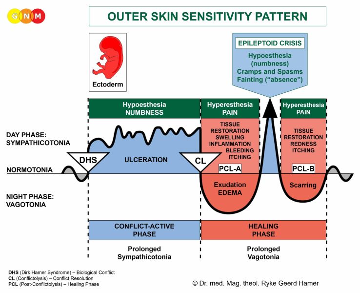
 DEVELOPMENT AND FUNCTION OF THE RENAL PELVIS AND URETERS: The renal pelvis and ureters represent the upper urinary tract. The renal pelvis receives urine from the kidney collecting tubules through their cup-shaped calyces. From there, urine flows into the ureters and further to the bladder and urethra (lower urinary tract) for elimination. The inner wall of the renal pelvis and ureters is endowed with smooth and striated muscles. Like the intestinal muscles that move the “food morsel” along the intestinal canal through peristaltic motion, the smooth muscles of the renal pelvis and ureters facilitate the flow of the “urine morsel”. The lining of the renal pelvis, including the renal calyces, and ureters consists of squamous epithelium, originates from the ectoderm and is therefore controlled from the cerebral cortex.
BIOLOGICAL CONFLICT: The biological conflict linked to the renal pelvis and the ureters is a male territorial marking conflict or a female marking conflict (see also bladder and urethra) depending on a person’s gender, laterality, and hormone status (see also Marking Constellation). A male territorial marking conflict refers to an unexpected invasion of the outer boundaries (male mammals mark the outer boundary of the territory with urine by hiking up their legs) whereas a female marking conflict relates to a breach of the inner boundaries (female mammals mark the inner boundary of their place by squatting). The female marking conflict is similar to an identity conflict, involving the rectum surface mucosa. This is why the brain relay of the renal pelvis, ureters, bladder and urethra is located next to the rectum relay (in the left temporal lobe).
A territorial marking conflict refers to an intrusion into one’s place (home, property), including the extended territory (neighborhood, village, city, country). Work-related marking conflicts are provoked, for example, through fights over a position or when a competitor moves into the professional terrain. Relationship-related marking conflicts concern members of the domain (spouse, children, parents, relatives, roommates, classmates, friends, visitors, neighbors, colleagues, teachers, supervisors) who are “crossing the line” or meddling in one’s business. Feeling controlled by a spouse, partner, or parent can evoke a marking conflict. An invasion of one’s private sphere also includes disrespect for one’s belongings. A man can suffer a territorial marking conflict, when another male is interested in his female or when his wife or girlfriend sleeps with someone else. Unwanted sex or sexual abuse can be perceived as an invasion of one’s intimate space. An assault against one’s beliefs, racist remarks, or harassment of any kind could prompt a marking conflict. Children experience the conflict at school, kindergarten, daycare, or on the playground, also, when a new sibling is born, when they have to share the room with a family member, or when they fight over a toy. Pets suffer marking conflicts when other animals (or humans) occupy their territory or when they are relocated.
CONFLICT-ACTIVE PHASE: ulceration in the lining of the renal pelvis, renal calyces and/or ureter(s) proportional to the degree and duration of conflict activity. The biological purpose of the cell loss is to enlarge the volume of the renal pelvis and to widen the ureter(s) to improve the urine flow in order to be better able to mark the territory.
HEALING PHASE: During the first part of the healing phase (PCL-A) the tissue loss is replenished through cell proliferation with swelling due to the edema (fluid accumulation) in the healing area. Healing symptoms are burning pain during urination (when the ureters are affected) and potentially blood in the urine (see also kidney parenchyma, bladder trigone, bladder mucosa, and prostate). Depending on the intensity of the conflict, the symptoms range from mild to severe. A large swelling can obstruct the affected ureter! An inflammation in the renal pelvis is called pyelitis. The Epileptoid Crisis manifests as acute pain with cramps or spasm (ureteric colic, kidney colic) if the surrounding striated muscles of the renal pelvis and/or ureters undergo the Epileptoid Crisis at the same time (see also kidney colic related to the kidney collecting tubules).
A “bacterial infection” in the renal pelvis or ureters indicates that the repair and scarring process (PCL-B) is assisted by bacteria. This is usually the case when the ulceration that occurred in the conflict-active phase reaches deep into the renal or ureteral tissue (see also “kidney infection” related to the kidney collecting tubules). Recurring “infections” point to conflict relapses triggered by setting on tracks that were established when the original marking conflict took place.
|


BLADDER
 DEVELOPMENT AND FUNCTION OF THE BLADDER TRIGONE: The bladder trigone is the triangular area between the openings of the ureters and the urethra. When the bladder muscle contracts, the trigone funnels urine that is temporarily stored in the bladder into the urethra. Equal to the intestinal cells that digest and absorb food, the biological function of the bladder trigone is to “digest” (secretory quality) proteins and “absorb” (absorptive quality) urine (similar to the kidney collecting tubules). The submucosa of the bladder trigone consists of intestinal cylinder epithelium, originates from the endoderm and is therefore controlled from the brainstem.
BIOLOGICAL CONFLICT: The biological conflict linked to the bladder trigone is an ugly, “dirty” morsel conflict (dirty business, dirty tricks, dirty sex, etc.) similar to a “shit conflict” related to the sigmoid colon and rectum submucosa.
CONFLICT-ACTIVE PHASE: Starting with the DHS, during the conflict-active phase cells in the bladder trigone proliferate proportionally to the intensity of the conflict. The biological purpose of the cell increase is to improve the ability to “digest” or “absorb” the “dirty morsel”. With prolonged conflict activity a flat (absorptive type) or cauliflower-shaped growth (secretory type) forms in the trigone. In conventional medicine, this is diagnosed as a bladder cancer (compare with “bladder cancer” related to the bladder mucosa). If the rate of cell division exceeds a certain limit, then the cancer is considered “malignant”; below that limit, the growth is regarded as “benign” or diagnosed as a bladder polyp (see also healing phase).
HEALING PHASE: Following the conflict resolution (CL), fungi or mycobacteria such as TB bacteria remove the cells that are no longer needed. This causes purulent tuberculous cystitis, a “bacterial bladder infection”.
Healing symptoms are pain due to the swelling, a cloudy urine, potentially blood in the urine (see also kidney parenchyma, renal pelvis and ureters, bladder mucosa, and prostate), and night sweats. Depending on the degree of the conflict-active phase, the symptoms range from mild to severe.
When fungi participate in the healing process, this causes “candida cystitis”, which becomes chronic when a person is in a hanging healing due to conflict relapses. Contrary to the claims of conventional medicine, the fungal “infection” in the endodermal(!) bladder trigone cannot “spread” to other areas in the urinary tract such as to the ureters, bladder or urethra (originating from the ectoderm) because fungi don’t cross the germ layer threshold!
If the required microbes are not available upon the resolution of the conflict, because they were destroyed through an overuse of antibiotics, the additional cells remain. Eventually, the growth becomes encapsulated with connective tissue. This is usually diagnosed as a bladder polyp or as a “benign cancer” (see also conflict-active phase).
|

 DEVELOPMENT AND FUNCTION OF THE BLADDER MUCOSA AND URETHRA: The bladder and urethra make up the lower urinary tract. In females, the bladder lies in front of the uterus; the urethra is positioned near the front wall of the vagina. In males, the urethra extends to the end of the penis and carries urine as well as semen during ejaculation; at the neck of the bladder, the urethra is surrounded by the prostate. The bladder is a hollow muscular organ where urine received from the renal pelvis and ureters is temporarily stored. Urine exits the bladder through the urethra. The inner wall of the urethra is endowed with smooth and striated muscles. Like the intestinal muscles that move the “food morsel” along the intestinal canal through peristaltic motion, the smooth muscles of the urethra facilitate the flow and elimination of the “urine morsel”. The lining of the bladder and urethra consists of squamous epithelium, originates from the ectoderm and is therefore controlled from the cerebral cortex.
BIOLOGICAL CONFLICT: The biological conflict linked to the bladder mucosa and urethra is a male territorial marking conflict or a female marking conflict (see renal pelvis and ureters), depending on a person’s gender, laterality, and hormone status (see also Marking Constellation).
CONFLICT-ACTIVE PHASE: ulceration in the bladder mucosa and/or in the lining of the urethra proportional to the degree and duration of conflict activity. The biological purpose of the cell loss is to enlarge the volume of the bladder and to widen the urethra to improve the urine flow in order to be better able to mark the territory.
HEALING PHASE: During the first part of the healing phase (PCL-A) the tissue loss is replenished through cell proliferation with swelling due to the edema (fluid accumulation) in the healing area. In conventional medicine, this might be diagnosed as a “bladder cancer” or urothelial carcinoma, also called transitional cell carcinoma (compare with bladder cancer related to the bladder trigone). Based on the Five Biological Laws, the new cells cannot be regarded as “cancer cells” since the cell increase is, in reality, a replenishing process. A small, wart-like growth in the lining of the urinary tract, including the renal pelvis and ureters, is referred to as a “urothelial papilloma” and usually considered as “benign”.
Healing symptoms are frequent urges to void with burning pain during urination and elimination of only small amounts of urine; there is potentially blood in the urine (see also kidney parenchyma, renal pelvis and ureters, bladder trigone, and prostate). Typical is also the feeling of constantly needing to urinate and of incomplete emptying of the bladder following urination, a condition termed bladder tenesmus (compare with rectal tenesmus). With water retention due to the SYNDROME the enlarged swelling might block the urine flow in the urethra. This is an acute medical situation! In this case, Dr. Hamer recommends a temporary bladder catheter (see also urinary tract obstruction in males caused by an enlarged prostate or a prostate tumor).
The Epileptoid Crisis manifests as acute pain with cramps or spasm if the surrounding striated muscles of the inner wall of the urethra undergoes the Epileptoid Crisis at the same time.
A urinary tract infection in the urethra (urethritis) or a bladder infection (cystitis) indicates that the repair and scarring process (PCL-B) is assisted by bacteria (see also UTI related to the ureters and “bladder infections” related to the bladder trigone and bladder muscle). This is usually the case when the ulceration that occurred in the conflict-active phase reaches deep into the urethral and bladder tissue. Recurring “bladder infections” point to conflict relapses triggered by setting on a track that was established when the original marking conflict took place.
Urethral gonorrhea is an inflammation of the mucous membrane of the urethra with discharge due to the activity of bacteria (Neisseria gonorrhoeae) during the healing process. If Chlamydia trachomatis bacteria are involved, this causes a so-called “chlamydia infection”; chlamydia bacteria are also involved in urethritis (chlamydia in the mouth relates to an oral conflict; in the rectum or anus to an identity conflict). Contrary to standard beliefs, gonorrhea or chlamydia cannot be sexually transmitted since the symptoms are already healing symptoms, explicitly, of a (territorial) marking conflict regarding the sexual space (see also sexual separation conflict and genital herpes). If the symptoms are less severe, the condition might be diagnosed as urethritis or cystitis. What is euphemistically termed “honeymoon cystitis” is caused by frequent and prolonged sexual intercourse. NOTE: In men, the urethra is also used for ejaculation. Hence, the Biological Special Program of the urethra corresponds also to an ejaculation conflict (see also ejaculatory ducts) as in “not being able, not being allowed, or not wanting to ejaculate”, for example, premature ejaculation.
Bladder warts are the result of a prolonged healing in the urinary bladder. Erroneously these harmless residues are interpreted as cancers. Bladder warts are quite common in cats and dogs (territorial marking conflict!).
|

 DEVELOPMENT AND FUNCTION OF THE INTERNAL BLADDER SPHINCTER: The internal bladder sphincter is a ring-shaped muscle located at the lower neck of the bladder. Its muscular mechanism involuntarily regulates the flow of urine from the bladder into the urethra. The external bladder sphincter at the lower end of the urethra provides a second means to control urine elimination. The internal bladder sphincter consists of smooth muscles, originates from the endoderm and is controlled from the midbrain.
BIOLOGICAL CONFLICT: The biological conflict linked to internal bladder sphincter is not being able to hold back urine, for example, because of incontinence. Urinary incontinence is one of the most frequent causes of conflicts following a prostate surgery.
CONFLICT-ACTIVE PHASE: hypertonus of the internal bladder sphincter. The biological purpose of the increased muscle tension is to facilitate holding urine in the bladder.
HEALING PHASE: The muscle tension goes back to normal. The Epileptoid Crisis presents as painful bladder spasms (see also spasms in the ureters, bladder muscle, bladder mucosa and urethra).
|

 DEVELOPMENT AND FUNCTION OF THE BLADDER MUSCLE AND EXTERNAL BLADDER SPHINCTER: The bladder is a hollow organ for storing urine. The bladder wall consists of muscles which contract during urination forcing urine out of the bladder into the urethra; at the same time, the two sphincters open to allow urine to be expelled. The external bladder sphincter surrounds the lower end of the urethra and is, in addition to the internal bladder sphincter, a second muscular mechanism that regulates the elimination of urine. The striated bladder muscle and external bladder sphincter derive from the new mesoderm and are controlled from the cerebral medulla and the motor cortex.
BIOLOGICAL CONFLICT: The biological conflict linked to the bladder muscle and external bladder sphincter is “not being able to sufficiently mark one’s place” (see also external rectal sphincter). The conflict typically occurs when a territorial marking conflict cannot be resolved for a long period of time. The bladder muscles also relate to a self-devaluation conflict, usually brought on by urinary incontinence.
CONFLICT-ACTIVE PHASE: cell loss (necrosis) of bladder muscle tissue (controlled from the cerebral medulla) and, proportional to the degree of conflict activity, increasing paralysis of the bladder muscle (controlled from the motor cortex). At the same time, the external bladder sphincter opens (no necrosis with sphincters!), which increases the urine flow in order to be better able to mark the territory.
Urinary incontinence, an involuntary outflow of urine, is a sign that a persistent marking conflict is still unresolved. Depending on the intensity of the conflict, the condition ranges from mild leaking (when coughing, sneezing, laughing) to uncontrollable wetting (see also fecal incontinence). A sudden urine outflow also occurs during the Epileptoid Crisis when the bladder sphincter opens. Incontinence often generates self-devaluation conflicts involving adjacent tissues such as the pubic bone or pelvic floor muscles. Hence, weak pelvic floor muscles don’t cause incontinence but are rather the result of continuing bladder-related self-devaluation conflicts; the same holds true to recurring “bladder infections”.
Bedwetting (nocturnal enuresis) is the unintentional voiding of urine during sleep. The involuntary urination takes place during the Epileptoid Crisis which typically occurs at night, that is, in vagotonia. With the brief sympathicotonic stress, the bladder sphincter opens causing the urine excretion. Persistent or chronic bedwetting indicates that the person has continual conflict relapses followed by the “nighttime accident”. Children suffer territorial marking conflicts with their siblings or schoolmates or when they are physically, verbally, or socially bullied. NOTE: A complete emptying of the bladder can happen in the course of any intense Epileptoid Crisis.
HEALING PHASE: During the healing phase the bladder muscle is reconstructed and the bladder sphincter closes. If bacteria assist healing, this causes a “bacterial bladder infection” (see also bladder trigone and bladder mucosa) with painful bladder spasms during the Epileptoid Crisis (see also spasms related to ureters, internal bladder sphincter, bladder and urethra).
 | ||||||||||||||||||||


