NOSE and SINUSES


DEVELOPMENT AND FUNCTION OF THE NASAL MUCOSA: The nasal cavity is divided into a right and left passageway that connect with the paranasal sinuses through small orifices. In the back they join with the nasopharynx and the mouth. Of the five senses (sight, smell, taste, touch, hearing) the olfactory sense is the oldest. In humans, it is the most powerful sense at birth. The sense of smell is to a large extent linked with the sense of taste. The mucosa covering the inside of the nose cleans and moistens the air before entering the lungs. The nasal mucosa consists of squamous epithelium, originates from the ectoderm and is therefore controlled from the cerebral cortex.

NOTE: The nasal cavities are no longer endowed with an endodermal submucosa. However, the epithelial nasal mucosa still contains residues of endodermal cells (“nasal glands”) that produce nasal mucus (see also paranasal sinuses).


BRAIN LEVEL: The nasal mucosa is controlled from the sensory cortex (part of the cerebral cortex). The mucosa of the right nasal cavity is controlled from the left side of the sensory cortex; the mucosa of the left nasal cavity is controlled from the right cortical hemisphere (deep basal). Hence, there is a cross-over correlation from the brain to the organ (see GNM diagram showing the sensory homunculus).
BIOLOGICAL CONFLICT: The biological conflict linked to the nasal mucosa is according to its function a scent conflict (see also paranasal sinuses and “scent morsel” conflict related to the nasopharynx). For animals, the conflict might be provoked by the scent of an approaching predator or the smell of poisonous fumes. For humans, the conflict translates into “smelling” trouble or a potential threat, for instance, “smelling” a competitor or an opponent at work, at school, at home, or in a relationship. The nasal mucosa corresponds also to a stink conflict. A stink conflict is experienced in real terms through an offending odor or unpleasant smell, but also if the particular smell is associated with danger. The exposure to cigarette smoke can, therefore, trigger the conflict for someone who believes that second-hand smoke causes lung cancer. In a transposed sense, a stink conflict relates to any situation that is perceived as “This stinks!” or “I am fed up with this!”. This might also concern an annoying person (a “pest”). It is a type of “separation conflict”.

NOTE: Whether the right or left nasal cavity is affected is determined by a person’s handedness and whether the conflict is mother/child or partner-related. A general “stink conflict” affects both sides.


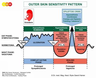
The Biological Special Program of the nasal mucosa follows the OUTER SKIN SENSITIVITY PATTERN with hyposensitivity during the conflict-active phase and the Epileptoid Crisis and hypersensitivity in the healing phase.           
CONFLICT-ACTIVE PHASE: ulceration of the nasal mucosa proportional to the degree and duration of conflict activity. The biological purpose of the cell loss is to widen the nasal passages in order to enhance the sense of smell (in Nature, smelling a predator or other potential dangers is essential for survival). Symptom: a dry nose due to the loss of nasal mucus-producing cells. During the conflict-active phase, the ulcers don't bleed. However, with a hanging conflict, they form crusts. 
HEALING PHASE: During the first part of the healing phase (PCL-A) the ulcerated area is replenished through cell proliferation. Healing symptoms are a stuffed up nose caused by the swelling of the nasal membrane, a reduced sense of taste and smell (compare with anosmia related to the olfactory nerves), nasal discharge to eliminate the remnants of the repair process, headaches because of the brain edema in the corresponding brain relay, elevated temperature or fever, and fatigue since the autonomic nervous system is in the “warm phase and in a prolonged state of rest (vagotonia). The shivers occur in the conflict-active “cold phase” as well as throughout the Epileptoid Crisis. Sneezing and nosebleeds are also a sign of the Epi-Crisis. In short, the healing phase of the nasal mucosa presents as the typical common cold. The degree of the symptoms is determined by the intensity of the conflict-active phase.

NOTE: All Epileptoid Crises that are controlled from the sensory, post-sensory, or pre-motor sensory cortex are accompanied by troubled circulation, dizzy spells, short disturbances of consciousness or a complete loss of consciousness (fainting or “absence”), depending on the intensity of the conflict. Another distinctive symptom is a drop of blood sugar caused by the excessive use of glucose by the brain cells (compare with hypoglycemia related to the islet cells of the pancreas).  

When the cold is accompanied or preceded by a sore throat, this indicates that the scent or stink conflict happened together with a conflict of not wanting to “swallow” a situation or accept what “stinks”. Coughing, related to the bronchi or larynx, reveals an additional territorial fear or scare-fright conflict. Typical for this conflict combination is unexpected distress at work, at school, or at home. As soon as the conflicts are resolved, the healing symptoms start all at once or in quick succession.

If a number of people have a cold at the same time, we can conclude that everyone who is affected had perceived a certain conflict situation the same way (troubles in daycare or kindergarten, poor marks for all students, an unfair teacher, arguments involving several family members, problems at the workplace) and is now in healing. In the northern hemisphere such collective “This stinks!”-conflicts are usually brought on at the beginning of the winter season – but only for those who “hate winter”. In spring, the same symptoms are referred to as the “seasonal flu”.

Conventional medicine claims that the cold or flu (see also influenza) are caused by viruses. However, to this day, the evidence of the existence of these alleged viruses has never been provided (details presented in the “Virus Mania” GNM video). Moreover, the symptoms of the cold and flu are healing symptoms, which highly questions the persistent claim that they are “contagious”.
Recurring or chronic cold symptoms occur when the scent or stink conflict is reactivated by setting on a conflict track such as a certain smell (food, perfume, flower, grass, cigarette smoke) or taste (milk, nuts, a spice), pet dander, pollen, mold, wind, rain, and so forth. In conventional medicine, this is usually interpreted as an “allergy”. People with pollen allergies might, in reality, be “allergic” to the cold symptoms (“This stinks!”) or to the “threat” of the “allergy season” resulting in common-cold symptoms (termed “allergic rhinitis”) each year. If the nasal congestion is accompanied by watery eyes (see conjunctivitis) then the “allergy” is called “hay fever”. In GNM terms, the combination of the symptoms indicates that the healing phases of a scent or stink conflict and a visual separation conflict (“I don’t want to see this!”) run concurrently.


On this CT scan we see the impact of a stink conflict in the area of the sensory cortex that controls the nasal mucosa of the left half of the nasal cavity (view the GNM diagram). For a right-handed person the conflict is associated with his/her mother or child; for a left-hander with a partner. The uneven, partly edematous ring of the Hamer Focus reveals that the person has already resolved the conflict and is now in the healing phase with symptoms of a cold.


DEVELOPMENT AND FUNCTION OF THE PARANASAL SINUSES: The paranasal sinuses are symmetrically arranged hollow, air-filled cavities lined by a mucous membrane. They are located behind the eyebrows (frontal sinuses), behind the nasal cavities (sphenoid sinuses), between the eyes and nose (ethmoid sinuses), and behind the cheekbones (maxillary sinuses). Their function is to moisten and warm the inhaled air and produce mucus that cleans the nasal passages. The paranasal sinuses mucosa consists of squamous epithelium, originates from the ectoderm and is therefore controlled from the cerebral cortex. Like the nasal cavities, the paranasal sinuses contain residues of endodermal cells (“paranasal glands”) that produce nasal mucus.

NOTE: The paranasal sinuses are the site from where the ectoderm (outer embryonic germ layer) emerged.


BRAIN LEVEL: The paranasal sinuses mucosa is controlled from the pre-motor sensory cortex (part of the cerebral cortex). The mucosa of the right sinuses is controlled from the left side of the cortex; the mucosa of the left sinuses is controlled from the right cortical hemisphere (fronto-basal). Hence, there is a cross-over correlation from the brain to the organ.

NOTE: The nasal mucosa is controlled from the sensory cortex.
BIOLOGICAL CONFLICT: The biological conflict linked to the paranasal sinuses is the same as the conflict related to the nasal mucosa, namely a scent conflict or stink conflict.


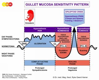
The Biological Special Program of the paranasal sinuses mucosa follows the GULLET MUCOSA SENSITIVITY PATTERN with hypersensitivity during the conflict-active phase and the Epileptoid Crisis and hyposensitivity in the healing phase.                               
CONFLICT-ACTIVE PHASE: ulceration in the mucosa of the paranasal sinuses proportional to the degree and duration of conflict activity. The biological purpose of the cell loss is to enhance the sense of smell. Symptom: mild to severe pain.

NOTE: Whether the mucosa of the right or left sinuses is affected is determined by a person’s handedness and whether the conflict is mother/child or partner-related. A general “stink conflict”  involves both sides. Which one of the paranasal sinuses is affected by the DHS is random.


This CT scan shows an active Hamer Focus with a sharp ring configuration on the right side of the pre-motor sensory cortex (view the GNM diagram) for the left paranasal sinuses, linked to a scent or stink conflict related to a partner if the person is left-handed; for a right-handed person the conflict is associated with his/her mother or child.
HEALING PHASE: During the first part of the healing phase (PCL-A) the tissue loss is replenished through cell proliferation. Healing symptoms are swelling of the sinus membrane due to the edema (fluid accumulation), nasal congestion, throbbing headaches (sinus headaches) and facial pain. The pain could last throughout the entire healing phase (in PCL-A and PCL-B the pain is not of a sensory nature but rather pressure pain). Concurrent water retention because of the SYNDROME enlarges the swelling and increases the pain.

An inflammation of the sinuses is called sinusitis. Recurring sinusitis indicates conflict relapses triggered by setting on a track that was established when the original stink conflict took place. The claim that sinusitis is caused by a “viral infection” is purely hypothetical.

NOTE: All Epileptoid Crises that are controlled from the sensory, post-sensory, or pre-motor sensory cortex are accompanied by troubled circulation, dizzy spells, short disturbances of consciousness or a complete loss of consciousness (fainting or “absence”), depending on the intensity of the conflict. Another distinctive symptom is a drop of blood sugar caused by the excessive use of glucose by the brain cells (compare with hypoglycemia related to the islet cells of the pancreas).  


Polyps in the paranasal sinuses are growths in the squamous epithelial sinus mucosa. They typically develop in the ethmoid and maxillary sinuses from where they grow into the nasal cavity (compare with nose polyps in the submucosa of the nasopharynx). With a hanging healing, that is, when the healing phase is continually interrupted by conflict relapses, the polyps can completely close the nasal passages.


DEVELOPMENT AND FUNCTION OF THE OLFACTORY NERVES: The olfactory nerves play a significant role in the sense of smell. They are composed of a collection of sensory nerve fibers (fila olfactoria) that extend down from the olfactory bulbs located at the frontal base of the cerebral cortex. Endowed with special receptor cells the olfactory nerves carry the olfactory signal from the mucosa at the roof of the nasal cavity to the olfactory bulbs. From there the information is transmitted to the brain where the smell is perceived on a conscious level. The olfactory nerves originate from the ectoderm and are controlled from the diencephalon.

BRAIN LEVEL: The olfactory nerves are controlled from the diencephalon (interbrain), which is located in the central part of the cerebrum just above the brainstem. The olfactory nerves in the left nasal cavity are controlled from the right side of the diencephalon; the olfactory nerves in the right nasal cavity are controlled from the left side (a right-handed female smells with the left nostril her child and with the right nostril her partner; for left-handers, it is reversed).

There is a cross-over correlation from the brain to the organ.
BIOLOGICAL CONFLICT: The biological conflict linked to the olfactory nerves is “not being able to smell something or someone” (in Nature this occurs when a female cannot smell a lost offspring) or, the opposite, “not wanting to smell something or someone”, for example, an overpowering stench or the odor of a rival.
CONFLICT-ACTIVE PHASE: functional loss of the olfactory nerves with the biological purpose to block the olfactory memory (equal to the short-term memory loss during conflict activity of a separation conflict) or the perception of the unwanted odor. The result is a reduced ability to smell the odor associated with the conflict (hyposmia; compare with hyperosmia) or a complete loss of smell (anosmia).

NOTE: The olfactory nerves belong to the group of organs that respond to the related conflict not with cell proliferation or cell loss but with hyperfunction (see also periosteum and thalamus) or functional loss (see Biological Special Programs of the inner ear (cochlea and vestibular organ), retina and vitreous body of the eyes, islet cells of the pancreas (alpha islet cells and beta islet cells), skeletal muscles).
HEALING PHASE: During the healing phase, the sense of smell is restored, shortly interrupted with a temporary loss of smell during the Epileptoid Crisis.  


This CT scan presents a Hamer Focus in PCL-A with fluid accumulation (brain edema) in the control center of the olfactory nerves (red arrows - view the GNM diagram), indicating that the related conflict has been resolved. With water retention due to an active abandonment or existence conflict involving the kidney collecting tubules (yellow arrow), the brain edema increases significantly.
HYPEROSMIA

Olfactory hypersensitivity (hyperosmia), an increased sensitivity to smell, relates biologically to the sensitivity of the original gullet.


BRAIN LEVEL: In the brainstem, the brain relays of the olfactory nerve (first cranial nerve) are evenly distributed over the control centers of the gastrointestinal tract.

The biological conflict linked to the primordial intestinal sensitivity is “not being able to sufficiently smell or identify a (food) morsel”.  The oversensitivity to smells occurs in the conflict-active phase. The biological purpose is to be better able to identify the “morsel” (in Nature this is vital for survival). During the healing phase the sense of smell returns to normal.


NOSE and SINUSES - TRANSLATIONS

<---- In these PDF documents you can enlarge images. If your browser cannot handle this desirable feature, then you will need to download and install the authentic Adobe Acrobat Reader

Click on the icon to download the PDF file

English Original
Catalan Translation
Dutch Translation

French Translation
Russian Translation
Serbian Translation

Spanish Translation

Turkish Translation